

Particle Concentration Measurement
Advanced Characterization of Extracellular Vesicles and Exosomes for Biomedical Research, Diagnostics and Biopharmaceutical Development
Extracellular vesicle (EV) analysis has rapidly emerged as one of the most dynamic and impactful fields in life science research. Once considered merely cellular debris, extracellular vesicles are now recognized as fundamental mediators of intercellular communication, carrying proteins, lipids, nucleic acids and metabolites that reflect the physiological state of their cells of origin. Their involvement in cancer biology, neurodegenerative diseases, cardiovascular disorders, immune regulation and regenerative medicine has positioned extracellular vesicles—and particularly exosomes—at the center of modern biomedical research.
As interest in liquid biopsy, precision medicine and cell-based therapies continues to grow, the ability to accurately characterize extracellular vesicles has become increasingly important. Researchers require analytical techniques capable not only of measuring particle size and concentration but also of distinguishing heterogeneous vesicle populations with high sensitivity and reproducibility.
Extracellular vesicle samples are inherently complex. Biological fluids such as plasma, serum, urine, cerebrospinal fluid or cell culture media contain mixed populations of vesicles differing in size, composition, membrane structure and biological origin. Conventional particle sizing techniques often struggle to resolve this complexity, making comprehensive characterization a significant analytical challenge.
Advanced extracellular vesicle analysis therefore relies on multiparametric approaches capable of combining particle size distribution, particle concentration and optical characterization at the single-particle level. This multidimensional information enables researchers to better understand vesicle heterogeneity, optimize isolation protocols and generate more reliable experimental data.
Whether supporting biomarker discovery, therapeutic development or fundamental biological research, extracellular vesicle characterization has become an essential component of modern nanobiotechnology.
What Are Extracellular Vesicles?
Extracellular vesicles (EVs) are membrane-bound nanoparticles naturally released by virtually all cell types into the extracellular environment. Acting as biological messengers, they transport molecular cargo between cells, influencing numerous physiological and pathological processes.
Unlike synthetic nanoparticles, extracellular vesicles are highly complex biological structures composed of lipid bilayers enclosing proteins, messenger RNA, microRNA, DNA fragments, enzymes and other bioactive molecules. Their molecular composition reflects the characteristics of the originating cell, making them valuable sources of biological information.
Extracellular vesicles are generally classified into several categories based on their biogenesis, although significant overlap exists between different populations.
Exosomes are among the most extensively studied extracellular vesicles. Typically ranging from approximately 30 to 150 nm in diameter, they originate from the endosomal pathway through the formation of multivesicular bodies before being secreted into the extracellular space.
Microvesicles are generally larger and are produced by direct outward budding of the plasma membrane. Apoptotic bodies, released during programmed cell death, represent an additional vesicle population with distinct biological characteristics.
Although these classifications remain useful, the International Society for Extracellular Vesicles (ISEV) recommends describing vesicles primarily according to measurable characteristics such as size, density, biochemical composition and cellular origin rather than assuming precise biogenesis pathways.
Why Extracellular Vesicle Analysis Matters
The scientific interest surrounding extracellular vesicles has increased dramatically because these nanoparticles participate in virtually every aspect of cell-to-cell communication.
Their biological cargo provides unique insight into cellular physiology, enabling researchers to investigate disease progression through minimally invasive biological samples.
In oncology, extracellular vesicles contribute to tumor progression, angiogenesis, metastasis and immune modulation. Their molecular content may serve as a valuable source of biomarkers for early cancer detection and therapeutic monitoring.
In neurology, extracellular vesicles are increasingly investigated for Alzheimer's disease, Parkinson's disease, multiple sclerosis and traumatic brain injury because they can transport disease-associated proteins across the blood-brain barrier.
Cardiovascular research has likewise demonstrated the involvement of extracellular vesicles in thrombosis, inflammation, endothelial dysfunction and myocardial injury.
Beyond diagnostics, extracellular vesicles are attracting considerable attention as potential therapeutic delivery systems. Their natural biocompatibility, low immunogenicity and ability to transport biological cargo make them promising candidates for targeted drug delivery and regenerative medicine.
As these applications continue to expand, robust extracellular vesicle characterization becomes essential for ensuring experimental reproducibility and translating laboratory discoveries into clinical practice.
Challenges in Extracellular Vesicle Characterization
Despite significant technological progress, extracellular vesicle analysis remains challenging due to the intrinsic complexity of biological samples.
Individual EV populations exhibit broad variability in size, refractive index, membrane composition and cargo content. Furthermore, extracellular vesicle preparations often contain protein aggregates, lipoproteins, cell debris and other nanoparticles that may interfere with accurate characterization.
No single analytical technique currently provides complete information regarding extracellular vesicle populations.
Researchers therefore frequently combine complementary analytical methods to obtain reliable characterization data.
The ability to distinguish different nanoparticle populations without extensive sample manipulation has consequently become one of the major goals in extracellular vesicle research.
Conventional Techniques for Extracellular Vesicle Analysis
Several analytical techniques are commonly employed to characterize extracellular vesicles.
Nanoparticle Tracking Analysis (NTA) provides particle size and concentration measurements by tracking Brownian motion of individual particles. While widely used, its performance may decrease when samples contain highly heterogeneous particle populations.
Dynamic Light Scattering (DLS) offers rapid average particle size estimation but is strongly influenced by larger particles, making complex biological samples difficult to interpret.
Transmission Electron Microscopy (TEM) provides exceptional morphological detail but requires extensive sample preparation and does not represent routine quantitative analysis.
Flow Cytometry enables phenotypic characterization of larger vesicles using fluorescent markers but remains limited for smaller extracellular vesicles without specialized instrumentation.
Resistive Pulse Sensing (RPS) and tunable nanopore technologies provide additional sizing capabilities but often require careful optimization depending on sample composition.
Because each technique measures different physical properties, combining complementary analytical information remains the preferred strategy for comprehensive extracellular vesicle characterization.
Multiparametric Extracellular Vesicle Analysis with SPES Technology
SPES technology developed by EOS Instruments, implemented in the Classizer™ ONE, introduces a multiparametric single-particle approach for the larger fraction of extracellular vesicle samples, from approximately 300 nm up to 20 µm, including large EVs, apoptotic bodies and non-vesicular particles such as cell debris and aggregates. Exosomes and other small EVs, typically below 200 nm, lie below the SPES detection range: their effective refractive index is close to that of the surrounding medium, and they are typically measured with techniques such as NTA, resistive pulse sensing or high-sensitivity flow cytometry. SPES complements these methods by characterizing larger vesicles and particles that are often under-represented in nanoscale measurements.
Rather than relying solely on particle diameter, SPES simultaneously measures particle size distribution, effective refractive index, particle concentration and optical characteristics, generating a multidimensional description of each detected particle.
This approach enables researchers to distinguish particle populations that would otherwise appear similar when evaluated exclusively by size measurements. In EV preparations, this can help separate large vesicles from non-vesicular particles, such as cell debris, protein aggregates or large lipoprotein particles, which differ in effective refractive index.
For extracellular vesicle research, this additional level of characterization can improve the interpretation of complex biological samples, support optimization of isolation protocols and facilitate comparison between different experimental conditions.
EOS CLOUDS visualization further enhances data interpretation by representing particle populations according to multiple optical parameters, helping researchers identify distinct subpopulations of large vesicles and non-vesicular particles within heterogeneous samples.
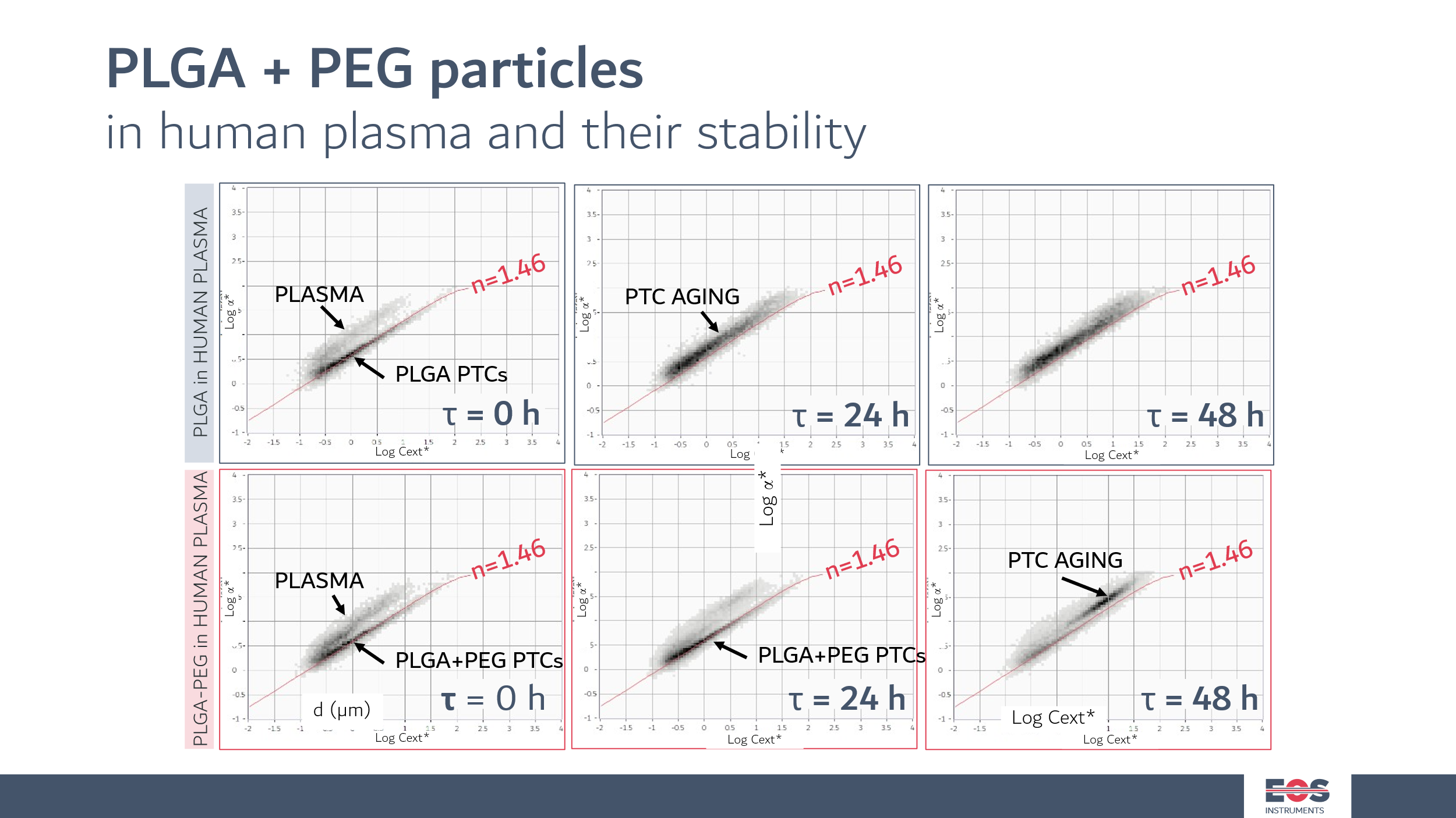
In figure, PLGA nanoparticles with high RI of 1.46 are measured alongside a blood sample (upper cloud). In the first row, SPES data of PLGA made without the use of a surfactant as a stabilizier. In the course of 48 h, the particles degrades and age. In the bottom row, PLGA particles stabilized with a surfactant. As it can be seen, they survive almost unalterated at the 24h timestamp. At 48 h, a population with bigger sizes and lower RI can be observed that can be interpreted as swelling of the particles.
Applications
Extracellular vesicle analysis supports numerous research and industrial applications, including:
Exosome research
Liquid biopsy development
Cancer biomarker discovery
Neurodegenerative disease research
Cardiovascular disease studies
Regenerative medicine
Stem cell research
Drug delivery systems
Vaccine research
Nanomedicine
Biopharmaceutical development
Academic research laboratories
Clinical translational research
For exosome research and other applications focused on small EVs, SPES is used to monitor large vesicles, aggregates and non-vesicular particles above approximately 300 nm, for example to check EV isolates for large contaminants, complementing nanoscale characterization techniques.
Conclusion
Extracellular vesicle analysis has become one of the most rapidly evolving areas of biomedical research. As the scientific community continues to recognize the diagnostic and therapeutic potential of extracellular vesicles, analytical technologies capable of accurately characterizing heterogeneous nanoparticle populations are becoming increasingly important.
While conventional techniques remain indispensable, modern extracellular vesicle research requires multidimensional characterization capable of integrating particle size, concentration and optical properties at the single-particle level.
SPES technology contributes to this analytical perspective for the larger vesicle fraction, enabling researchers to investigate large extracellular vesicles and non-vesicular particles with greater depth while supporting biomarker discovery, therapeutic development and the growing field of precision medicine.
Related posts
