

Particle Concentration Measurement
Advanced Liposome Characterization for Drug Delivery, Nanomedicine and Pharmaceutical Development
Liposome characterization is a fundamental analytical process in modern pharmaceutical development, nanomedicine and drug delivery research. Since their introduction as versatile lipid-based delivery systems, liposomes have become one of the most widely investigated nanoparticle platforms for improving drug stability, enhancing therapeutic efficacy and enabling targeted delivery of active pharmaceutical ingredients.
Today, liposomal formulations are used in numerous clinical applications, including cancer therapy, vaccines, gene delivery and controlled drug release. More recently, the rapid expansion of lipid nanoparticle technologies has further emphasized the importance of accurately characterizing lipid-based nanocarriers throughout development and manufacturing.
The therapeutic performance of liposomes depends on multiple physicochemical properties. Particle size, size distribution, concentration, membrane integrity, colloidal stability and particle heterogeneity all influence circulation time, cellular uptake, biodistribution and drug release kinetics. Even relatively small changes in these parameters may significantly affect formulation quality and clinical performance.
For this reason, liposome characterization extends far beyond measuring average particle diameter. Modern analytical workflows increasingly combine particle sizing, concentration measurements and optical characterization to obtain a multidimensional understanding of liposomal formulations.
Comprehensive liposome characterization supports formulation optimization, manufacturing process development, stability studies, quality control and regulatory compliance while helping researchers develop safer, more effective lipid-based therapeutics.
What Are Liposomes?
Liposomes are spherical vesicles composed of one or more phospholipid bilayers surrounding an aqueous core. Because their structure closely resembles biological cell membranes, liposomes are highly biocompatible and capable of encapsulating both hydrophilic and hydrophobic therapeutic compounds.
Hydrophilic molecules can be incorporated within the internal aqueous compartment, while hydrophobic drugs become embedded inside the phospholipid bilayer. This unique structural versatility has made liposomes attractive delivery vehicles for a wide variety of pharmaceutical applications.
Depending on their preparation method, liposomes may exhibit different structural characteristics, including:
Small Unilamellar Vesicles (SUVs)
Large Unilamellar Vesicles (LUVs)
Giant Unilamellar Vesicles (GUVs)
Multilamellar Vesicles (MLVs)
Each population presents distinct size distributions, encapsulation efficiencies and biological behaviors, making accurate characterization essential during formulation development.
Why Liposome Characterization Matters
The physicochemical properties of liposomes directly determine their therapeutic performance.
Particle size strongly influences circulation half-life, tissue penetration and cellular internalization. Smaller liposomes may circulate longer within the bloodstream, while larger particles may be removed more rapidly by the reticuloendothelial system.
Particle size distribution also plays an important role. Highly homogeneous formulations generally exhibit greater reproducibility, whereas broad distributions may indicate inconsistent manufacturing processes or particle instability.
Liposome concentration affects dosing accuracy and manufacturing consistency. Accurate concentration measurements are particularly important during scale-up and quality control.
Long-term formulation stability represents another critical aspect. Liposomes may undergo aggregation, fusion, leakage or structural degradation during storage, potentially altering encapsulation efficiency and therapeutic activity.
Comprehensive characterization therefore enables researchers to optimize formulations while ensuring reproducibility throughout product development and manufacturing.
Key Parameters for Liposome Characterization
Modern liposome characterization evaluates numerous physicochemical properties simultaneously.
Among the most important analytical parameters are:
Particle size
Particle size distribution
Particle concentration
Lamellarity
Encapsulation efficiency
Membrane integrity
Colloidal stability
Surface charge (Zeta Potential)
Population heterogeneity
Together, these parameters provide a detailed description of liposomal formulations and support formulation optimization across the entire development process.
Conventional Techniques for Liposome Characterization
Several analytical techniques are routinely employed to characterize liposomal formulations.
Dynamic Light Scattering (DLS) remains one of the most commonly used methods for determining average particle size and size distribution. Although rapid and easy to perform, DLS provides ensemble measurements that may be strongly influenced by larger particles or aggregates.
Nanoparticle Tracking Analysis (NTA) measures individual particle motion to estimate both particle size and concentration. This technique offers valuable single-particle information but may become less reliable for highly concentrated or heterogeneous samples.
Cryogenic Transmission Electron Microscopy (Cryo-TEM) provides detailed structural visualization of liposomes, allowing direct observation of membrane architecture and particle morphology. However, it requires specialized instrumentation and is generally unsuitable for routine quality control.
Additional characterization methods include Size Exclusion Chromatography (SEC), Asymmetric Flow Field-Flow Fractionation (AF4), Resistive Pulse Sensing (RPS) and Zeta Potential measurements, each providing complementary information regarding different formulation attributes.
Because liposomes represent highly heterogeneous nanoparticle systems, no individual technique fully characterizes every relevant physicochemical property.
Beyond Particle Size: Multiparametric Liposome Characterization
Average particle size alone rarely provides sufficient information to understand complex liposomal formulations.
Different liposome populations may exhibit similar diameters while differing substantially in membrane composition, internal structure, lipid packing or encapsulated cargo. These differences may significantly influence biological performance despite remaining undetected through conventional size measurements.
Multiparametric characterization can help address this limitation by integrating particle size distribution with additional particle properties, including concentration, optical behavior and effective refractive index.
This broader analytical perspective allows researchers to detect formulation heterogeneity, monitor subtle stability changes and better evaluate manufacturing consistency.
As liposomal therapeutics become increasingly sophisticated, multiparametric analysis is emerging as an important complement to conventional particle sizing techniques.
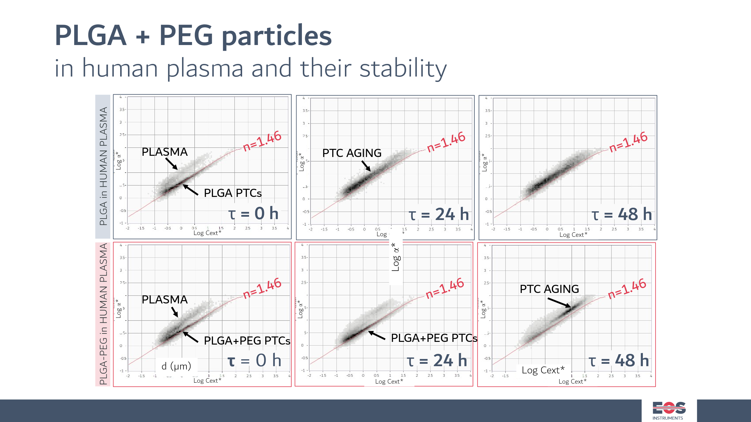
In figure, PLGA nanoparticles with high RI of 1.46 are measured alongside a blood sample (upper cloud). In the first row, SPES data of PLGA made without the use of a surfactant as a stabilizier. In the course of 48 h, the particles degrades and age. In the bottom row, PLGA particles stabilized with a surfactant. As it can be seen, they survive almost unalterated at the 24h timestamp. At 48 h, a population with bigger sizes and lower RI can be observed that can be interpreted as swelling of the particles.
Liposome Characterization with SPES Technology
SPES technology developed by EOS Instruments, implemented in the Classizer™ ONE, provides an advanced multiparametric approach for characterizing liposomal formulations at the single-particle level, focusing on aggregates, multilamellar liposomes and other particles above approximately 300–400 nm.
Rather than measuring particle size alone, SPES simultaneously evaluates particle size distribution, particle concentration, effective refractive index and optical characteristics for each detected particle. This multidimensional characterization enables researchers to identify heterogeneous particle populations that may remain hidden when relying exclusively on average particle size measurements. Because small unilamellar liposomes consist mostly of water enclosed by a thin lipid bilayer, their effective refractive index is very close to that of the surrounding medium. Their main nanoscale population is therefore best measured with techniques such as DLS or NTA, while SPES characterizes the larger and optically denser fraction of the formulation.
The capability to distinguish particles according to both size and optical properties provides valuable insight into formulation complexity, aggregation phenomena and manufacturing consistency.
EOS CLOUDS visualization further enhances interpretation by displaying multidimensional particle populations in an intuitive graphical representation, facilitating the identification of distinct liposome subpopulations, such as aggregates, fusion products or multilamellar vesicles, and improving analytical confidence.
This analytical strategy supports formulation development, process optimization, quality control and advanced research involving lipid-based drug delivery systems.
Applications
Liposome characterization supports a wide range of pharmaceutical and biomedical applications, including:
Cancer therapeutics
Nanomedicine
Vaccine development
mRNA delivery systems
Lipid nanoparticle (LNP) research
Gene therapy
Controlled drug release
Pharmaceutical formulation development
Process development
Manufacturing optimization
Academic research
Biotechnology
Translational medicine
For lipid nanoparticles, mRNA delivery systems and small unilamellar liposomes, whose primary population lies below the SPES detection range, SPES is used to monitor aggregates, oversized particles and larger lipid structures above approximately 300–400 nm, complementing nanoscale sizing techniques in stability studies and quality control.
Conclusion
Liposome characterization has become an essential analytical discipline supporting the development of modern lipid-based therapeutics. As pharmaceutical formulations become increasingly complex, comprehensive characterization of particle populations is critical for ensuring product quality, stability and therapeutic performance.
While conventional particle sizing techniques remain indispensable, advanced formulations require a broader analytical perspective capable of integrating particle size, concentration and optical characterization into a single workflow.
SPES technology by EOS Instruments complements established liposome characterization techniques through multiparametric single-particle analysis of aggregates and larger lipid structures, providing researchers with deeper insight into formulation heterogeneity while supporting innovation in nanomedicine, drug delivery and pharmaceutical development.
Related posts
